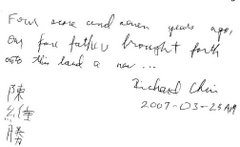
We received an email from Jane Qiu today about her recently published article in the September 2009 issue of Nature Biotechnology. Jane had interviewed Richard in 2007 about his stem cell therapy experience in China which resulted in an article published by the Lancet Neurology in February 2008.
The Nature Biotechnology article which is entitled "Trading on Hope" is a followup to her investigation of "the thriving business of selling stem cell transplants as cure-alls for debilitating diseases." Jane has graciously made available a copy of her article to us.
Please click on the link, TradingonhopeNatureBiotech9-09.pdf, here.
Furthermore, Jonathan Kimmelman, who holds a PhD in Molecular Biophysics and Biochemistry from Yale University and is currently with the Biomedical Ethics Unit Faculty at the McGill University in Montreal, Canada, commented on Jane's article in his blog (Lost in Translation) post of September 23, 2009.
Saturday, October 10, 2009
Friday, October 2, 2009
Perchance to dream
On our last visit with Dr. Susan Perlman at UCLA, Richard had complained about how he was suffering from parasomnia. He has experienced, on a regular basis, night terrors, sleep talking, bruxism and periodic leg movement. He has even fallen out of bed several times as a result of his nightmares. Needless to say, the quality of his sleep was less than satisfactory in general.
After consulting with her colleague, Dr. Perlman prescribed melatonin, which is OTC, to address the issue. We are happy to report that after taking this drug for a few months, the symptoms have abated. He is now sleeping more soundly which in turn translates to more energy during the day.
I continue to be gratified by how Dr. Perlman is able to tackle problems that plague Richard and other MJDers by using existing drugs or supplements. For example, melatonin is used mainly to help with circadian rhythm disorders like jet-lag disorders. Similarly, she suggested Fluoxetine (trade name Prozac) to help Richard with swallowing and choking problems and that too has worked well in the last two years. I hope and believe that there are many more drugs and supplements out there that one day will be shown to be equally effective in treating various MJD symptoms.
Wednesday, September 30, 2009
The latest research on MJD/SCA3
Here's an article from PloS ONE where researchers are trying to understand the behavior and movement of material of the nucleus (center) of the cell of someone who has MJD. Ataxin-3, I believe is the mutated genetic material within an MJDer's body cells that causes the ataxia. With a clearer picture as to how the "bad stuff" gets transported into the cells, the researchers can then think of and experiment with different ways in preventing that from happening.
It is a very technical article. I am nowhere close to even understanding 10% of what is written. Nevertheless, for those who are interested in learning more about this subject, here is the article in its entirety:
PLoS ONE. 2009; 4(6): e5834.
Published online 2009 June 8. doi: 10.1371/journal.pone.0005834.
PMCID: PMC2688764
Copyright Macedo-Ribeiro et al. This is an open-access article distributed under the terms of the Creative Commons Attribution License, which permits unrestricted use, distribution, and reproduction in any medium, provided the original author and source are credited.
Nucleocytoplasmic Shuttling Activity of Ataxin-3
Sandra Macedo-Ribeiro,#1* Luísa Cortes,#2 Patrícia Maciel,3 and Ana Luísa Carvalho2,4*
1IBMC-Instituto de Biologia Molecular e Celular, Universidade do Porto, Porto, Portugal
2Center for Neuroscience and Cell Biology (CNC), University of Coimbra, Coimbra, Portugal
3Life and Health Sciences Research Institute (ICVS), School of Health Sciences, University of Minho, Braga, Portugal
4Department of Zoology, University of Coimbra, Coimbra, Portugal
David C. Rubinsztein, Editor
University of Cambridge, United Kingdom
#Contributed equally.
* E-mail:sribeiro@ibmc.up.pt (SMR);Email: alc@cnc.cj.uc.pt (ALC)
Conceived and designed the experiments: SMR PM ALC. Performed the experiments: LC ALC. Analyzed the data: SMR LC ALC. Contributed reagents/materials/analysis tools: PM. Wrote the paper: SMR LC ALC.
Received March 4, 2009; Accepted May 8, 2009.
Top
>Abstract
Introduction
Methods
Results and Discussion
References
Abstract
Spinocerebellar ataxia type-3, also known as Machado-Joseph Disease (MJD), is one of many inherited neurodegenerative disorders caused by polyglutamine-encoding CAG repeat expansions in otherwise unrelated genes. Disease protein misfolding and aggregation, often within the nucleus of affected neurons, characterize polyglutamine disorders. Several evidences have implicated the nucleus as the primary site of pathogenesis for MJD. However, the molecular determinants for the nucleocytoplasmic transport of human ataxin-3 (Atx3), the protein which is mutated in patients with MJD, are not characterized.
In order to characterize the nuclear shuttling activity of Atx3, we performed yeast nuclear import assays and found that Atx3 is actively imported into the nucleus, by means of a classical nuclear localizing sequence formed by a cluster of lysine and arginine residues. On the other hand, when active nuclear export was inhibited using leptomycin B, a specific inhibitor of the nuclear export receptor CRM1, both endogenous Atx3 and transfected GFP-Atx3 accumulated inside the nucleus of a subpopulation of COS-7 cells, whereas both proteins are normally predominant in the cytoplasm.
Additionally, using a Rev(1.4)-GFP nuclear export assay, we performed an extensive analysis of six putative aliphatic nuclear export motifs identified in Atx3 amino acid sequence. Although none of the tested peptide sequences were found to drive nuclear export when isolated, we have successfully mapped the region of Atx3 responsible for its CRM1-independent nuclear export activity. Curiously, the N-terminal Josephin domain alone is exported into the cytoplasm, but the nuclear export activity of Atx3 is significantly enhanced in a longer construct that is truncated after the two ubiquitin interaction motifs, upstream from the polyQ tract.
Our data show that Atx3 is actively imported to and exported from the cell nucleus, and that its nuclear export activity is dependent on a motif located at its N-terminal region. Since pathological Atx3 aggregates in the nucleus of affected neurons in MJD, and there is in vivo evidence that nuclear localization of Atx3 is required for the manifestation of symptoms in MJD, defects in the nucleocytoplasmic shuttling activity of the protein may be involved in the nuclear accumulation and aggregation of expanded Atx3.
Top
Abstract
>Introduction
Methods
Results and Discussion
References
Introduction
Machado-Joseph disease (MJD), or spinocerebellar ataxia type 3, is the most common dominantly inherited ataxia worldwide and it is caused by a polyglutamine (polyQ) expansion in ataxin-3 (Atx3), a polyubiquitin-binding protein [1] with ubiquitin protease activity [2]. Full-length Atx3 contains an N-terminal Josephin domain (JD), the conserved catalytic module, two ubiquitin interacting motifs (UIMs), an expandable polyQ stretch, and a short variable tail that might contain a third UIM depending on the splice variant [3]. In normal individuals the size of the polymorphic glutamine repeat can range between 14 and 40 while in MJD patients the polyQ repeat is expanded to 53 or more glutamines [4]. Human Atx3 is ubiquitously expressed and displays a complex subcellular distribution involving both the cytoplasm and the nucleus, depending on cell type [5], [6], [7]. Even though its physiological role is still not clearly established, Atx3 was shown to be a cysteine protease with the ability to cleave polyubiquitin chains with more than four ubiquitins, independently of the polyglutamine tract [2]. This enzymatic activity of Atx3 has been correlated with its ability to mitigate polyQ-induced neurodegeneration in a Drosophila model [8], with its involvement in aggresome formation [9] and in the degradation of misfolded proteins [10]. Furthermore, it was also shown to bind to histones [11] and to chromatin [12] indicating that Atx3 displays not only cytoplasmic but also nuclear functions.
As for most other polyQ diseases, conformational changes imparted by the expanded polyQ tract lead to the formation of neuronal intranuclear inclusions (NIIs) [13] and may contribute to pathogenesis by affecting gene expression [14] or by disrupting nuclear organization and function [15]. In MJD patients specific brain regions are affected such as the cerebellum and brainstem, with prominent cell loss in the pontine and dentate nuclei [6], [16], [17]. It is becoming clear that although polyQ tracts themselves are toxic, the sequence and structure of the proteins carrying the polyQ tracts have important roles in defining the course and specificity of the disease. Those sequences determine subcellular localization, and specify interactions with other macromolecules within the cell, strongly determining the differences in the specificity of neuronal degeneration characteristic of polyQ disorders [18], [19], [20].
A contentious question has been whether polyQ-induced pathogenesis is primarily activated in the cytoplasm or in the cell nucleus. In fact predominantly nuclear inclusions have been found in SCA1, SCA2, SCA7, SCA17, DRPLA, SBMA, and Huntington's Disease (HD) patients [21], although cytoplasmic inclusions have also been identified in affected brain regions in SCA2 [22] and HD [23]. Evidence from HD transgenic mice shows that both nuclear and cytoplasmic exon-1 huntingtin might contribute to disease progression [23], [24], [25]. Nuclear environment has been shown to favor toxicity, pathology and aggregation as evidenced by nuclear targeting of polyQ peptides [26], even when inserted into ectopic protein contexts [27].
Specific nuclear localization sequences (NLS) have been identified in proteins carrying the expanded polyQ tracts, such as ataxin-1 [28], [29] and ataxin-7 [30], and nuclear-associated mechanisms are being implicated in neuropathogenesis [13], [31]. Similarly, nuclear export sequences (NES) have been found in ataxin-7 [32] and huntingtin [33] and it was shown that polyQ expansion impairs efficient nuclear export of these polyQ-containing proteins [32], [34].
Recently, it was demonstrated in vivo that adding an exogenous NLS to Atx3(148Q) increases the severity of the phenotype and induces earlier death in transgenic mouse models [35]. Accordingly, adding an exogenous NES to Atx3(148Q) drives the expanded protein out of the nucleus and prevents the manifestation of a phenotype [35]. This suggests that defects on the nucleocytoplasmic shuttling activity of the expanded protein might be correlated with pathology and neuronal specificity. Moreover, in other transgenic models of Machado-Joseph disease there is accumulation of the expanded Atx3 protein in the nucleus of affected neurons [36], [37], [38]. However, the molecular determinants for the nucleocytoplasmic transport of Atx3 are not characterized. In order to gain further insights into the function of Atx3 and into the disease-specific mechanisms of neurodegeneration in MJD, we have set as our goal the identification of the determinants of Atx3 nucleocytoplasmic transport.
Nuclear targeting was analyzed in vivo using the yeast system developed by Rhee et al. [39] and we found that Atx3 is actively imported into the nucleus, by means of a classical NLS located in its C-terminal region. Furthermore, using an in vivo nuclear export assay we show that Atx3 is actively exported from the nucleus and mapped this export activity to its N-terminal domain.
Top
Abstract
Introduction
>Methods
Results and Discussion
References
Methods
Yeast nuclear import assay
The yeast nuclear import assay was performed as described previously [39]. The cDNA encoding the MJD1.a isoform of ataxin-3 (AAB33571), containing 28 CAG repeats, was amplified by PCR using the pEGFP-C1-ataxin-3(28Q) construct (kindly provided by Dr. Henry Paulson) as template and using the primers 5′tcccccgggcatggtgagcaagggcg3′ and 5′gcgtcgacttatgtcagataaagtgtgaag3′, which introduced SmaI and SalI recognition sites at the 5′ and 3′ ends of the amplified DNA fragment, respectively. The cDNA encoding ataxin-3(28Q) was cloned into the SmaI and SalI sites of the plasmid for the yeast nuclear import assay (pNIA, kindly provided by Vitaly Citovsky [39]), and named pNIA-GFP-Atx3. The pNIA-GFP-Atx3R282T and pNIA-GFP-Atx3R282A constructs were obtained by site-directed mutagenesis (QuickChange site-directed mutagenesis kit, Stratagene).
The pNIA constructs, encoding triple fusion proteins comprising bacterial LexA, yeast Gal4p activation domain (Gal4AD), and the tested protein, were transformed using the lithium acetate method [40] into Saccharomyces cerevisiae L40 strain, which contains the reporter genes HIS3 and lacZ with upstream LexA operators. After transformation, yeasts were plated on selective medium deficient for tryptophan, to select for transformed cells. Transformed yeasts were then plated on selective medium lacking both tryptophan and histidine, and supplemented with 100 mM 3-amino-1,2,4-triazole (3AT; Sigma), an inhibitor of the His3p enzyme, and growth was evaluated. Additionally, transformed yeasts were grown in tryptophan-deficient liquid medium for quantitative determination of β-galactosidase activity [41]. For the enzymatic assay, cells were disrupted, and the β -galactosidase substrate o-nitrophenyl-β-D-galactopyranoside (ONPG) was added in excess. The reaction occurred at 30°C and was stopped by raising the pH to 11. The optical density of the reaction product was measured at 420 nm (OD420), and the β-galactosidase activity was calculated according to the following equation: βunits = 1,000×OD420/t×V×OD600, where OD420 is the optical density at 420 nm of the sample measured after the incubation of yeast cell lysate with ONPG, t is the time of incubation (in minutes) of the yeast cell lysate with ONPG, V is the volume of the sample used in the assay (in milliliters), and OD600 is the optical density at 600 nm of the yeast cell culture at the start of the assay.
Cell culture, transfection and leptomycin B treatment
HEK293 and COS-7 cells were grown and maintained in Dulbecco's modified Eagle's medium-high glucose (DMEM-HG; Sigma) supplemented with 10% (vol/vol) heat-inactivated fetal bovine serum (FBS; Biochrom KG) and with 100 U of penicillin and 100 µg of streptomycin (Sigma) per ml in a 5% CO2 humidified atmosphere at 37°C.
One day prior to transfection, COS-7 cells were seeded onto glass coverslips on a 12-well plate, at a subconfluent density. Transfection experiments were performed using Lipofectamine reagent (Invitrogen) according to the manufacturer's instructions, using 1 µg of plasmid DNA per well. The cells were incubated for 48 hours to allow gene expression.
Where indicated, the cells were incubated with 20 ng/ml leptomycin B (Sigma) in DMEM-HG supplemented with 10% FBS for 3 hours prior to fixation.
Rev(1.4)-GFP nuclear export assay
To assess the strength of the nuclear export activity of different fragments of Atx3 protein, or of full-length Atx3, the Rev(1.4)-GFP nuclear export assay, described previously by Henderson and Eleftheriou [42], was used. This assay is based on the manipulation of the Rev protein shuttling cycle, and tests for the ability of functional nuclear export sequences to promote the nuclear export activity of the Rev(1.4)-GFP fusion protein, which is composed of a NES-deficient mutant of the HIV-1 Rev protein and GFP.
Potential Atx3 NES signals were identified by consensus to the ΦX1–3ΦX2–3ΦXΦ motif (Φ indicates a large hydrophobic residue, and X indicates any amino acid), and using the prediction algorithm NetNES (http://www.cbs.dtu.dk/databases/NESbase-1.0 [43]), and fused to Rev(1.4)-GFP fusion protein (the plasmid encoding Rev(1.4)-GFP was kindly provided by Beric R. Henderson). The putative sequences that were assayed include: NES1, residues 76-SIQVISNALKVWGLELILF-94; NES2, 133-LNSLLT-138; NES3, 142-LISDTYLALFLAQLQQE-158; NES4, 174-ADQLLQMIRV-183; NES5, 209-LERVLE-214; and NES6, 222-LDEDEEDLQRALALSRQEIDME-243. Furthermore, full-length Atx3 (28Q and 84Q) or partial domains of Atx3 (the Josephin domain and the domains comprising amino acids 1–263 and 183–263) were also fused to Rev(1.4)-GFP fusion protein.
COS-7 cells were transfected with pRev(1.4)-GFP (negative control) or its derivative plasmids containing either the NES of the HIV-1 Rev protein (positive control) or each of the sequences to be tested. Forty-eight hours post-transfection, all cell samples were treated with 10 µg/ml cycloheximide (CHX) to ensure that any cytoplasmic GFP fluorescence resulted only from the nuclear export of GFP fusion proteins and not from de novo protein synthesis. Simultaneously, part of the cell samples were also treated for 3 hours with 5 µg/ml actinomycin D (ActD), which is known to specifically block the nuclear import of Rev protein by a mechanism not yet elucidated [42]. The remaining cell samples were treated with CHX, ActD and 20 ng/ml of leptomycin B for 3 hours. The subcellular localization of each GFP fusion protein was determined in at least 200 cells per experimental condition from three independent experiments.
Fluorescence microscopy
Endogenous Atx3 was detected, by immunocytochemistry, with anti-MJD antibody (1[ratio]10000), kindly provided by H. Paulson, and visualized using a secondary antibody labeled with Alexa 488 (1[ratio]1000, Invitrogen). For fluorescence analysis of GFP fusion proteins, cells were washed with phosphate-buffered saline (PBS), fixed with 4% paraformaldehyde: 4% sacarose for 15 min, and rinsed with PBS. The coverslips were then inverted and mounted on glass slides with Vectashield mounting medium (Vector Laboratories). The cell nucleus was stained with Hoescht 33342 (0,5 µg/ml, Molecular Probes). Fluorescence observations were performed using a Zeiss Axiovert 200 fluorescence microscope, coupled to a digital photographic camera (Axiocam HRM). Confocal microscopy was performed using a Zeiss LSM 510 Meta system.
Top
Abstract
Introduction
Methods
>Results and Discussion
References
Results and Discussion
Ataxin-3 has a mixed cytoplasmic and nuclear distribution
Ataxin-3 is an ubiquitous protein that is found both in the cytoplasm [44] and in the nucleus [5], [7]. However, upon expansion of the polyQ tract the protein forms insoluble inclusions predominantly located inside the nucleus of the affected cells [6]. Interestingly, the localization of the protein inside the cell is critically dependent on the cell type [5], [7]. Immunostaining of HEK293T cells with an anti-Atx3 antibody (kindly provided by H. Paulson [6]) showed that endogenous Atx3 is predominantly located in the nucleus (Fig. 1aFigure 1), while its distribution is more homogeneous in COS-7 cells (Fig. 1cFigure 1). Interestingly, when both cell lines were transiently transfected with Atx3 N-terminally tagged with GFP (GFP-Atx3 (28Q), Figs. 1e and 1gFigure 1), Atx3 was found predominantly in the cytoplasm of both cell types, in agreement with previous data [5], [6], [45].
Figure 1
Figure 1
Figure 1
Ataxin-3 can shuttle between the nucleus and the cytoplasm.
In order to determine whether Atx3 can be actively transported across the nuclear membrane we analyzed the effect of leptomycin B, a specific covalent inhibitor of the nuclear export factor CRM1/exportin [46], [47], on the subcellular distribution of both endogenous and overexpressed Atx3 in HEK293 and COS-7 cell lines. In eukaryotic cells, nuclear export of proteins is frequently mediated by this nuclear export factor, which binds to nuclear export signals (NES) on cargo molecules. If Atx3 is a nuclear shuttling protein, interfering with its putative nuclear export would be expected to modify Atx3 subcellular localization by increasing the proportion of the protein localized in the nucleus. Indeed, after cell treatment with leptomycin B both endogenous Atx3 (Figs. 1b and fFigure 1) and transfected GFP-Atx3 (28Q) (Figs. 1d, hFigure 1) accumulate in the nucleus of a subpopulation of cells, suggesting that Atx3 exits the cell nucleus by active transport at least partially dependent on the CRM1/exportin pathway. In COS-7 cells the nuclear accumulation of GFP-Atx3(28Q) could be observed in 26.5±4.5% of cells after leptomycin B treatment, whereas in untreated cells only 4.8±2.4% of cells showed nuclear accumulation of the protein. Nuclear export dependent on CRM1 receptor has also been demonstrated for other proteins containing expandable polyglutamine tracts such as ataxin-7 [32] and huntingtin [33]. In agreement with what is observed for Atx3 in the context of the full-length protein, leptomycin B treatment of cultured cells tranfected with huntingtin lead to a partial nuclear accumulation of the protein corresponding to a 10% increase in nuclear fluorescence of huntingtin [33].
Ataxin-3 contains a functional nuclear localization signal
Translocation of macromolecules larger than 40–60 kDa in and out of the nucleus is an active, energy-dependent process that is mediated by specific sequence motifs: nuclear localization signals (NLS) and nuclear export signals (NES). “Classical” examples of NLSs are the highly basic motifs originally found in the simian virus 40 (SV40) large T antigen and in nucleoplasmin, although several sequences differing from those basic ones have also been shown to function as NLS [48], [49], [50]. In “classical” nuclear import, importin-α recognizes and binds the target proteins in the cytoplasm, mediating their transport across the nuclear pore complex after formation of a ternary complex with importin-β [48], although some NLS-containing cargoes can be directly recognized by importin-β without the need of the adaptor protein. Within importin-α, the major NLS binding pocket contains a series of negatively charged residues that form salt bridges with the basic residues within the NLS sequences [51]. A putative NLS has been identified within the amino acid sequence of Atx3 [5], which is conserved among Atx3 proteins from diverse species (Fig. 2Figure 2).
Figure 2
Figure 2
Figure 2
Human ataxin-3 and its closest homologues contain conserved nuclear import sequences.
In order to test the functionality of the identified NLS sequence, we used a pNIA vector-based genetic one-hybrid system that allows identification of nuclear proteins in yeast cells [39]. This assay is based on the functional outcome of the nuclear import of the tested protein, which, if it reaches the yeast cell nucleus, allows specific induction of a reporter gene. The advantage of this approach is allowing the quantitative evaluation of the strength of the nucleocytoplasmic shuttling signals, independently of the cellular model. This system has been consistently used for mutational analysis of nuclear proteins to delineate and characterize the NLS [52] and for testing the integrity of the nuclear pore complex permeability barrier [53]. The rationale for this assay is the expression in the yeast S. cerevisiae cells of the test protein fused to a modified LexA DNA binding domain (DBD) and the Gal4 transcriptional activation domain (AD). This transcription based assay relies on the ability of a functional NLS to allow the chimera to enter the yeast nucleus and activate transcription of a LexA responsive β-galactosidase or HIS3 reporter gene. In the absence of a functional NLS, the fusion protein is not efficiently imported into the nucleus, and is unable to activate transcription. As a result, this assay provides a simple measure of NLS function based either on the quantitative determination of β-galactosidase activity or on the qualitative analysis of yeast growth in a medium lacking histidine. This system is not limited to the identification of yeast NLSs, as the nuclear import apparatus is highly conserved between yeast and higher eukaryotic cells [49], [50].
pNIA-GFP and pNIA-SV40NLS were used as controls for the nuclear import assay. pNIA-GFP encodes the fusion protein mLexA-Gal4AD-GFP and was used as negative control because it is not imported into the nucleus, resulting in minimal expression of the two reporter genes, whereas pNIA-SV40NLS encodes the fusion protein mLexA-SV40NLS-Gal4AD-GFP which, due to the presence of the SV40NLS, is actively imported into the nucleus, leading to high activity of both lacZ and HIS3 genes. The cDNA for ataxin-3 (with 28 glutamines) was cloned in the pNIA vector, and to ensure that the resulting fusion protein had a molecular weight not compatible with simple diffusion across the nuclear pore, GFP was inserted between Gal4AD and Atx3. When expressed in yeast, the pNIA-GFP-Atx3 fusion protein induced growth on histidine-deficient medium (data not shown), suggesting that Atx3 was actively imported into the nucleus. To confirm this result we performed a quantitative β-galactosidase assay, in liquid culture of yeast cells expressing the different constructs (Fig. 3Figure 3). The results obtained show that pNIA-GFPAtx3 induced levels of β-galactosidase activity significantly higher than the levels obtained for pNIA-GFP, a finding which confirms that Atx3 is actively imported into the nucleus of yeast cells.
Figure 3
Figure 3
Figure 3
Evaluation of the nuclear import capacity of ataxin-3 protein in yeast.
To determine whether the proposed NLS of Atx3 was indeed responsible for the translocation of the fusion protein into the nucleus of yeast cells, we mutated the conserved arginine residue within the putative NLS sequence into a threonine residue (pNIA-GFP-Atx3R282T). This mutation is a typical mutation performed in the analysis of conserved NLS sequences since it changes a basic residue for a neutral residue without modifying its polar character [28], and has also been shown to disrupt the function of the NLS identified in ataxin-1 and ataxin-7 [28], [30]. As shown in Figure 3Figure 3, the mutation results in the reduction the β-galactosidase activity to levels similar to the negative control (pNIA-GFP), indicating that the mutation greatly impairs Atx3 nuclear import in yeast. The mutation of the same arginine residue to an alanine residue (pNIA-GFP-Atx3R282A) was also tested, and resulted in a reduction on the nuclear accumulation of the fusion protein (Fig. 3Figure 3). These data show that interference with the putative NLS disrupts the nuclear import ability of Atx3.
The nuclear import activity of Atx3 was further confirmed in mammalian cells by comparing the subcellular localization of GFP-Atx3(28Q)R282T with GFP-Atx3(28Q) (Fig. 4Figure 4). When transfected in COS-7 cells, both constructs localized mainly in the cytoplasm of the cells. Because our data show that Atx3 can also be exported from the nucleus and that this export is at least partially mediated by the CRM1 pathway (see above), the similar localization of wild-type Atx3 and the R282T mutant might be due to the presence of competitive nuclear export signals. Therefore, in order to investigate if there is a difference between the nuclear shuttling ability of the wild-type protein and the R232T mutant in COS-7 cells, we incubated the cells with leptomycin B, thereby at least partially inhibiting nuclear export, and determined their subcellular re-localization. As shown in Fig. 1Figure 1 (panels g and h), GFP-Atx3(28Q) accumulates in the nucleus of a subpopulation of cells in the presence of leptomycin B. When GFP-Atx3(28Q)R282T is expressed in COS-7 cells this nuclear accumulation is not observed (Fig. 4Figure 4), indicating that the identified NLS sequence is also responsible for driving Atx3 into the nucleus of mammalian cells. Therefore, we conclude that Atx3 contains a basic NLS sequence that is functional and promotes its active import into the cell nucleus.
Figure 4
Figure 4
Figure 4
Evaluation of the nuclear import ability of ataxin-3 in mammalian cells.
The nuclear export of ataxin-3 is mediated by CRM1-dependent and -independent pathways
Since the nuclear export of Atx3 was partially inhibited in the presence of leptomycin B (Figs. 1Figure 1, ,4),4Figure 4), we analyzed the nuclear export activity of Atx3 using the Rev(1.4)-GFP nuclear export assay [42]. This assay is based on the expression of fusion proteins consisting of Rev(1.4), an export-defective HIV-1 Rev protein mutant, the sequence to be tested, and GFP. The nuclear export functionality of the sequence to be tested is evaluated by analyzing its capacity to promote nuclear export of the Rev(1.4)-GFP fusion protein. The Rev(1.4)-GFP fusion protein, which was used as a negative control for this assay, was localized exclusively in the nucleus of COS-7 cells, presenting a clear nucleolar accumulation (Fig. 5bFigure 5). Rev(1.4)-NES–GFP, a fusion protein that contains the Rev NES, which was used as a positive control for this assay, was localized in the nucleus and the cytoplasm of transfected cells (Fig. 5bFigure 5). The number of cells presenting exclusively cytoplasmic localization was enhanced following actinomycin D (ActD) treatment (Fig. 5bFigure 5), which blocks the nuclear import mediated by the Rev NLS [42], whereas the cytoplasmic localization of Rev(1.4)-NES-GFP was completely blocked by addition of leptomycin B (Fig. 5bFigure 5), as expected for a protein whose nuclear export is dependent on CRM1.
Figure 5
Figure 5
Figure 5
The nuclear export of ataxin-3 is mediated by the protein N-terminal domain.
Full-length Atx3 containing 28 glutamine residues [Atx3(28Q)], when fused to Rev(1.4)-GFP, was driven into the nucleus, where it formed punctuate structures resembling the insoluble nuclear inclusions (Fig. 5bFigure 5). The morphology of these structures is clearly distinct from the nucleolar accumulation of fluorescence observed for Rev(1.4)-GFP (Fig. 5bFigure 5). We also tested full-length Atx3 containing 84 glutamine residues [Atx3(84Q), Fig. 5bFigure 5], which formed punctuate structures, presumably aggregates, in the nucleus of transfected cells. These structures were not sensitive to the blockade of nuclear import of the Rev fusion protein using ActD. In fact, this result is in agreement with previous observations that Atx3 has a tendency to oligomerize independently of the expanded polyQ tract [54], [55], [56] and that the nuclear environment promotes protein misfolding and aggregation [57]. Curiously, it was observed that overexpression of ataxin-7 in cell culture models induced the nuclear localization of the protein and formation of large protein “accumulations” in ~30% of the cells, independently of the polyglutamine tract size [30].
The nuclear aggregation of full-length Atx3 when fused to Rev(1.4)-GFP, independently of the extent of the polyQ tract, presumably triggered by the strong Rev NLS, hampered the investigation of the nuclear export activity of full-length Atx3 using this system. Therefore, we prepared constructs encoding various domains of Atx3 fused to Rev(1.4)-GFP, all lacking the polyglutamine tract and the endogenous NLS (Fig. 5aFigure 5). The fusion protein consisting of the Josephin domain of Atx3 [JD-(1–182)] fused to Rev(1.4)-GFP showed a distribution between the cell nucleus and cytoplasm in 46% of the cells after cell treatment with ActD (Fig. 5bFigure 5). We also tested the cellular localization of the fusion protein consisting of the Josephin domain followed by the segment of Atx3 that contains the UIMs [Atx3(1–263)], fused to Rev(1.4)-GFP [Rev1.4-Atx3(1–263)-GFP]. This protein also displayed a nuclear and cytoplasmic localization, and the number of cells showing a mixed distribution of the protein increased to 78% after cell treatment with ActD (Fig. 5bFigure 5). The nuclear export activities of JD(1–182) and Atx3(1–263) were not inhibited by the CRM1 export inhibitor leptomycin B (+LMB).
These results suggest that the Josephin domain can mediate the nuclear export of the fusion proteins, which requires the context of the Josephin domain plus a sequence downstream this catalytic region. However, this UIM-containing segment alone [Rev(1.4)-Atx3(183–263)-GFP, Fig. 5bFigure 5] was not sufficient to promote nuclear export of the fusion protein. Finally, we asked whether the nuclear export of the Atx3(1–263) fragment was dependent on the ubiquitin binding capacity of its UIMs. It has been shown, in a cell-based assay of polyQ aggregation, that recruitment of non-expanded Atx3 into nuclear aggregates is mediated by its UIMs [58]. Therefore, we mutated the two UIMs in the Atx3(1–263) fragment in order to compromise their functionality [Atx3(1–263)(S236A,S256A)]. This protein had the same pattern of cellular distribution as the wild-type Atx3(1–263) fragment (Fig. 5bFigure 5), indicating that functionality of the UIMs is not required for the increased nuclear export activity observed for this region of Atx3.
Our results clearly demonstrate that nuclear export of Atx3 is dependent on a complex Atx3 motif, located in the N-terminal portion of the protein, which requires the context of the Josephin domain plus the ubiquitin-interacting motifs. Taken together, these data indicate either that nuclear export of Atx3 is mediated by a nuclear export receptor that recognizes a properly folded conformational motif and/or that multiple export pathways contribute to the overall nuclear export of full-length Atx3. Interestingly, a nuclear export receptor, exportin 7, has been recently described, which recognizes nuclear export signals that include conformation-dependent recognition motifs, rather than short linear sequences [59].
Since we observed nuclear accumulation of Atx3 in a population of COS-7 cells when the CRM1 transporter was inhibited with leptomycin B (Figs. 1Figure 1 and and4),4Figure 4), we looked for leucine-rich nuclear export signal (NES) sequences within Atx3 primary structure that might match the consensus NES sequence for the CRM1-dependent export [60], using the NetNES predictor (http://www.cbs.dtu.dk/databases/NESbase-1.0 [43]). We found 6 putative NES sequences, of which 4 are present within the Josephin domain (Fig. 2Figure 2, NES1-NES4), one is located between the Josephin domain and the first ubiquitin interaction domain (NES5) and the last one (NES6) corresponds to the first ubiquitin interaction motif of the protein (Fig. 2Figure 2). We have fused the putative NESs identified in Atx3 with Rev(1.4)-GFP, and tested whether the presence of these sequences can induce the translocation of Rev(1.4)-GFP from the cell nucleus. However, the isolated NES sequences failed to induce nuclear export when inserted into the Rev(1.4)-GFP vector (Table 1). The observation that in this system none of the tested sequences showed nuclear export activity can result from the fact that these sequences do not function as active leucine-rich nuclear export signals, but could also be a consequence of testing the sequences isolated from their overall protein context. For example, the isolated NES of Rev binds to CRM1 much more weakly than does the full-length Rev protein [61], implying that an NES may require flanking sequences to adopt the conformation needed for CRM1 binding. Another possibility is that each isolated sequence is not strong enough to drive detectable nuclear export of Rev1.4-GFP, which has a very strong NLS, and that several NESs work in concert to achieve efficient export of Atx3. In fact, recent studies show that most NESs bind to CRM1 with relatively low affinity, since high-affinity NES binding to CRM1 impairs the efficient release of export complexes from the nuclear pore complex [60].
Table 1
Table 1
Analysis of the nuclear export activity of putative NES sequences within ataxin-3 primary structure.
The nuclear export of GFP-Atx3(28Q) was partially inhibited by leptomycin B (Figs. 1Figure 1, ,4),4Figure 4), suggesting that a CRM1-dependent pathway is involved in the nuclear export of Atx3. On the other hand, using the Rev(1.4)-GFP fusion system, which contains a strong NLS, we detected nuclear export activity of the N-terminal portion of Atx3 (Josephin domain and UIMs) that was not sensitive to leptomycin B (Fig. 5bFigure 5). This supports the possibility that, in resemblance to what has been found for huntingtin [33], [34], CRM1-dependent and -independent nuclear export mechanisms coexist to cooperate in determining the subcellular distribution of Atx3. In the context of the full-length protein, one of these pathways may be dominant when compared to the other. Similar to that of Atx3 and huntingtin, the nuclear export of other proteins, such as α-catenin [62], receptor-interacting protein 3 (RIP3) [63] and the African Swine Fever Virus p37 protein [64], has been shown to be mediated by both CRM1-dependent and –independent pathways.
Conclusion
A correlation between the nuclear environment and protein aggregation in MJD models has been pinpointed by studies using Atx3 with an exogenously added NLS that demonstrated in situ that the nuclear environment drives aggregation of both expanded and non-expanded Atx3 [57], [65]. Recent in vivo studies have shown that adding an exogenous NES does not only suppress the formation of NIIs almost completely, but also seems to prevent the aggregation of Atx3 [35]. Furthermore, live-cell imaging studies showed that expanded polyQ tracts slow the dynamics of intact Atx3, and that the export of expanded Atx3 is less efficient than the export of normal Atx3 [66], suggesting a defect on the nucleocytoplasmic shuttling activity of the expanded protein. In fact, a recent study using a Drosophila system to screen for genetic modifiers of Atx3 neurodegeneration identified as a suppressor of Atx3-mediated toxicity, among others, the gene encoding the nuclear export protein Embargoed, the orthologue of human CRM1 [67]. In order to further understand the link between nuclear localization, aggregation and toxicity of expanded Atx3, it is essential to identify the mechanisms behind the intracellular dynamics of the normal protein. In this study, we have confirmed the functionality of the putative Atx3 NLS in yeast and mammalian cells, and detected CRM1-dependent and –independent pathways that mediate the nuclear export of Atx3. Our data show that the CRM1-independent nuclear export of Atx3 is mediated by the N-terminal region of the protein and is critically dependent on a three-dimensional motif whose integrity is compromised when the Josephin domain is physically separated from the UIMs. Future identification of the nuclear transporter(s) responsible for this pathway, will pave the way to determining how the subcellular localization of Atx3 is regulated in physiological or pathological conditions.
Acknowledgments
We are grateful to Henry Paulson for providing the pEGFP-C1-ataxin-3(28Q) plasmid, as well as the anti-ataxin-3 antibody, to Vitaly Citovsky for providing the pNIA vector and the Saccharomyces cerevisiae L40 strain, and to Beric R. Henderson for providing the Rev(1.4)-GFP and the Rev(1.4)-NES-GFP plasmids. We thank Ana Eulálio and Maria C. Pedroso de Lima for helpful advice and discussions.
Footnotes
Competing Interests: The authors have declared that no competing interests exist.
Funding: This work was supported by a grant from the Portuguese Foundation for Science and Technology (POCI/SAU-MMO/60156/2004) and by Crioestaminal/Associacao Viver a Ciencia. LC was a recipient of a post-doctoral fellowship from the Portuguese Foundation for Science and Technology (SFRH/BPD/20686/2004/22). The funders had no role in study design, data collection and analysis, decision to publish, or preparation of the manuscript.
Top
Abstract
Introduction
Methods
Results and Discussion
References
References
1.
Chai Y, Berke SS, Cohen RE, Paulson HL. Poly-ubiquitin binding by the polyglutamine disease protein ataxin-3 links its normal function to protein surveillance pathways. J Biol Chem. 2004;279:3605–3611. [PubMed]
2.
Burnett B, Li F, Pittman RN. The polyglutamine neurodegenerative protein ataxin-3 binds polyubiquitylated proteins and has ubiquitin protease activity. Hum Mol Genet. 2003;12:3195–3205. [PubMed]
3.
Masino L, Musi V, Menon RP, Fusi P, Kelly G, et al. Domain architecture of the polyglutamine protein ataxin-3: a globular domain followed by a flexible tail. FEBS Lett. 2003;549:21–25. [PubMed]
4.
Maciel P, Costa MC, Ferro A, Rousseau M, Santos CS, et al. Improvement in the molecular diagnosis of Machado-Joseph disease. Arch Neurol. 2001;58:1821–1827. [PubMed]
5.
Tait D, Riccio M, Sittler A, Scherzinger E, Santi S, et al. Ataxin-3 is transported into the nucleus and associates with the nuclear matrix. Hum Mol Genet. 1998;7:991–997. [PubMed]
6.
Paulson HL, Perez MK, Trottier Y, Trojanowski JQ, Subramony SH, et al. Intranuclear inclusions of expanded polyglutamine protein in spinocerebellar ataxia type 3. Neuron. 1997;19:333–344. [PubMed]
7.
Trottier Y, Cancel G, An-Gourfinkel I, Lutz Y, Weber C, et al. Heterogeneous intracellular localization and expression of ataxin-3. Neurobiol Dis. 1998;5:335–347. [PubMed]
8.
Warrick JM, Morabito LM, Bilen J, Gordesky-Gold B, Faust LZ, et al. Ataxin-3 suppresses polyglutamine neurodegeneration in Drosophila by a ubiquitin-associated mechanism. Mol Cell. 2005;18:37–48. [PubMed]
9.
Burnett BG, Pittman RN. The polyglutamine neurodegenerative protein ataxin 3 regulates aggresome formation. Proc Natl Acad Sci U S A. 2005;102:4330–4335. [PubMed]
10.
Wang Q, Li L, Ye Y. Regulation of retrotranslocation by p97-associated deubiquitinating enzyme ataxin-3. J Cell Biol. 2006;174:963–971. [PubMed]
11.
Li F, Macfarlan T, Pittman RN, Chakravarti D. Ataxin-3 is a histone-binding protein with two independent transcriptional corepressor activities. J Biol Chem. 2002;277:45004–45012. [PubMed]
12.
Evert BO, Araujo J, Vieira-Saecker AM, de Vos RA, Harendza S, et al. Ataxin-3 represses transcription via chromatin binding, interaction with histone deacetylase 3, and histone deacetylation. J Neurosci. 2006;26:11474–11486. [PubMed]
13.
Ross CA, Wood JD, Schilling G, Peters MF, Nucifora FC, Jr, et al. Polyglutamine pathogenesis. Philos Trans R Soc Lond B Biol Sci. 1999;354:1005–1011. [PubMed]
14.
Zoghbi HY, Orr HT. Glutamine repeats and neurodegeneration. Annu Rev Neurosci. 2000;23:217–247. [PubMed]
15.
Sun J, Xu H, Negi S, Subramony SH, Hebert MD. Differential effects of polyglutamine proteins on nuclear organization and artificial reporter splicing. J Neurosci Res. 2007;85:2306–2317. [PubMed]
16.
Sequeiros J, Coutinho P. Epidemiology and clinical aspects of Machado-Joseph disease. Adv Neurol. 1993;61:139–153. [PubMed]
17.
Schmidt T, Landwehrmeyer GB, Schmitt I, Trottier Y, Auburger G, et al. An isoform of ataxin-3 accumulates in the nucleus of neuronal cells in affected brain regions of SCA3 patients. Brain Pathol. 1998;8:669–679. [PubMed]
18.
Orr HT. Beyond the Qs in the polyglutamine diseases. Genes Dev. 2001;15:925–932. [PubMed]
19.
Paulson H. Polyglutamine neurodegeneration: minding your Ps and Qs. Nat Med. 2003;9:825–826. [PubMed]
20.
Lim J, Crespo-Barreto J, Jafar-Nejad P, Bowman AB, Richman R, et al. Opposing effects of polyglutamine expansion on native protein complexes contribute to SCA1. Nature. 2008
21.
Schols L, Bauer P, Schmidt T, Schulte T, Riess O. Autosomal dominant cerebellar ataxias: clinical features, genetics, and pathogenesis. Lancet Neurol. 2004;3:291–304. [PubMed]
22.
Huynh DP, Figueroa K, Hoang N, Pulst SM. Nuclear localization or inclusion body formation of ataxin-2 are not necessary for SCA2 pathogenesis in mouse or human. Nat Genet. 2000;26:44–50. [PubMed]
23.
DiFiglia M. Huntingtin fragments that aggregate go their separate ways. Mol Cell. 2002;10:224–225. [PubMed]
24.
Schilling G, Savonenko AV, Klevytska A, Morton JL, Tucker SM, et al. Nuclear-targeting of mutant huntingtin fragments produces Huntington's disease-like phenotypes in transgenic mice. Hum Mol Genet. 2004;13:1599–1610. [PubMed]
25.
Benn CL, Landles C, Li H, Strand AD, Woodman B, et al. Contribution of nuclear and extranuclear polyQ to neurological phenotypes in mouse models of Huntington's disease. Hum Mol Genet. 2005;14:3065–3078. [PubMed]
26.
Yang W, Dunlap JR, Andrews RB, Wetzel R. Aggregated polyglutamine peptides delivered to nuclei are toxic to mammalian cells. Hum Mol Genet. 2002;11:2905–2917. [PubMed]
27.
Jackson WS, Tallaksen-Greene SJ, Albin RL, Detloff PJ. Nucleocytoplasmic transport signals affect the age at onset of abnormalities in knock-in mice expressing polyglutamine within an ectopic protein context. Hum Mol Genet. 2003;12:1621–1629. [PubMed]
28.
Klement IA, Skinner PJ, Kaytor MD, Yi H, Hersch SM, et al. Ataxin-1 nuclear localization and aggregation: role in polyglutamine-induced disease in SCA1 transgenic mice. Cell. 1998;95:41–53. [PubMed]
29.
Irwin S, Vandelft M, Pinchev D, Howell JL, Graczyk J, et al. RNA association and nucleocytoplasmic shuttling by ataxin-1. J Cell Sci. 2005;118:233–242. [PubMed]
30.
Kaytor MD, Duvick LA, Skinner PJ, Koob MD, Ranum LP, et al. Nuclear localization of the spinocerebellar ataxia type 7 protein, ataxin-7. Hum Mol Genet. 1999;8:1657–1664. [PubMed]
31.
Orr HT. Qs in the nucleus. Neuron. 2001;31:875–876. [PubMed]
32.
Taylor J, Grote SK, Xia J, Vandelft M, Graczyk J, et al. Ataxin-7 can export from the nucleus via a conserved exportin-dependent signal. J Biol Chem. 2006;281:2730–2739. [PubMed]
33.
Xia J, Lee DH, Taylor J, Vandelft M, Truant R. Huntingtin contains a highly conserved nuclear export signal. Hum Mol Genet. 2003;12:1393–1403. [PubMed]
34.
Cornett J, Cao F, Wang CE, Ross CA, Bates GP, et al. Polyglutamine expansion of huntingtin impairs its nuclear export. Nat Genet. 2005;37:198–204. [PubMed]
35.
Bichelmeier U, Schmidt T, Hubener J, Boy J, Ruttiger L, et al. Nuclear localization of ataxin-3 is required for the manifestation of symptoms in SCA3: in vivo evidence. J Neurosci. 2007;27:7418–7428. [PubMed]
36.
Alves S, Regulier E, Nascimento-Ferreira I, Hassig R, Dufour N, et al. Striatal and nigral pathology in a lentiviral rat model of Machado-Joseph disease. Hum Mol Genet. 2008;17:2071–2083. [PubMed]
37.
Chen X, Tang TS, Tu H, Nelson O, Pook M, et al. Deranged calcium signaling and neurodegeneration in spinocerebellar ataxia type 3. J Neurosci. 2008;28:12713–12724. [PubMed]
38.
Goti D, Katzen SM, Mez J, Kurtis N, Kiluk J, et al. A mutant ataxin-3 putative-cleavage fragment in brains of Machado-Joseph disease patients and transgenic mice is cytotoxic above a critical concentration. J Neurosci. 2004;24:10266–10279. [PubMed]
39.
Rhee Y, Gurel F, Gafni Y, Dingwall C, Citovsky V. A genetic system for detection of protein nuclear import and export. Nat Biotechnol. 2000;18:433–437. [PubMed]
40.
Kaiser C, Michaelis S, Mitchell A. Methods in yeast genetics. Cold Spring Harbor, NY: Cold Spring Harbor Laboratory Press; 1994.
41.
Stachel SE, An G, Flores C, Nester EW. A Tn3 lacZ transposon for the random generation of beta-galactosidase gene fusions: application to the analysis of gene expression in Agrobacterium. Embo J. 1985;4:891–898. [PubMed]
42.
Henderson BR, Eleftheriou A. A comparison of the activity, sequence specificity, and CRM1-dependence of different nuclear export signals. Exp Cell Res. 2000;256:213–224. [PubMed]
43.
la Cour T, Gupta R, Rapacki K, Skriver K, Poulsen FM, et al. NESbase version 1.0: a database of nuclear export signals. Nucleic Acids Res. 2003;31:393–396. [PubMed]
44.
Paulson HL, Das SS, Crino PB, Perez MK, Patel SC, et al. Machado-Joseph disease gene product is a cytoplasmic protein widely expressed in brain. Ann Neurol. 1997;41:453–462. [PubMed]
45.
Chai Y, Koppenhafer SL, Shoesmith SJ, Perez MK, Paulson HL. Evidence for proteasome involvement in polyglutamine disease: localization to nuclear inclusions in SCA3/MJD and suppression of polyglutamine aggregation in vitro. Hum Mol Genet. 1999;8:673–682. [PubMed]
46.
Kudo N, Khochbin S, Nishi K, Kitano K, Yanagida M, et al. Molecular cloning and cell cycle-dependent expression of mammalian CRM1, a protein involved in nuclear export of proteins. J Biol Chem. 1997;272:29742–29751. [PubMed]
47.
Kudo N, Wolff B, Sekimoto T, Schreiner EP, Yoneda Y, et al. Leptomycin B inhibition of signal-mediated nuclear export by direct binding to CRM1. Exp Cell Res. 1998;242:540–547. [PubMed]
48.
Lange A, Mills RE, Lange CJ, Stewart M, Devine SE, et al. Classical nuclear localization signals: definition, function, and interaction with importin alpha. J Biol Chem. 2007;282:5101–5105. [PubMed]
49.
Pemberton LF, Paschal BM. Mechanisms of receptor-mediated nuclear import and nuclear export. Traffic. 2005;6:187–198. [PubMed]
50.
Sorokin AV, Kim ER, Ovchinnikov LP. Nucleocytoplasmic transport of proteins. Biochemistry (Mosc). 2007;72:1439–1457. [PubMed]
51.
Terry LJ, Shows EB, Wente SR. Crossing the nuclear envelope: hierarchical regulation of nucleocytoplasmic transport. Science. 2007;318:1412–1416. [PubMed]
52.
Yang B, Zhu W, Johnson LB, White FF. The virulence factor AvrXa7 of Xanthomonas oryzae pv. oryzae is a type III secretion pathway-dependent nuclear-localized double-stranded DNA-binding protein. Proc Natl Acad Sci U S A. 2000;97:9807–9812. [PubMed]
53.
Patel SS, Belmont BJ, Sante JM, Rexach MF. Natively unfolded nucleoporins gate protein diffusion across the nuclear pore complex. Cell. 2007;129:83–96. [PubMed]
54.
Chow MK, Paulson HL, Bottomley SP. Destabilization of a non-pathological variant of ataxin-3 results in fibrillogenesis via a partially folded intermediate: a model for misfolding in polyglutamine disease. J Mol Biol. 2004;335:333–341. [PubMed]
55.
Gales L, Cortes L, Almeida C, Melo CV, do Carmo Costa M, et al. Towards a structural understanding of the fibrillization pathway in Machado-Joseph's disease: trapping early oligomers of non-expanded ataxin-3. J Mol Biol. 2005;353:642–654. [PubMed]
56.
Masino L, Nicastro G, Menon RP, Dal Piaz F, Calder L, et al. Characterization of the structure and the amyloidogenic properties of the Josephin domain of the polyglutamine-containing protein ataxin-3. J Mol Biol. 2004;344:1021–1035. [PubMed]
57.
Perez MK, Paulson HL, Pittman RN. Ataxin-3 with an altered conformation that exposes the polyglutamine domain is associated with the nuclear matrix. Hum Mol Genet. 1999;8:2377–2385. [PubMed]
58.
Donaldson KM, Li W, Ching KA, Batalov S, Tsai CC, et al. Ubiquitin-mediated sequestration of normal cellular proteins into polyglutamine aggregates. Proc Natl Acad Sci U S A. 2003;100:8892–8897. [PubMed]
59.
Mingot JM, Bohnsack MT, Jakle U, Gorlich D. Exportin 7 defines a novel general nuclear export pathway. Embo J. 2004;23:3227–3236. [PubMed]
60.
Kutay U, Guttinger S. Leucine-rich nuclear-export signals: born to be weak. Trends Cell Biol. 2005;15:121–124. [PubMed]
61.
Paraskeva E, Izaurralde E, Bischoff FR, Huber J, Kutay U, et al. CRM1-mediated recycling of snurportin 1 to the cytoplasm. J Cell Biol. 1999;145:255–264. [PubMed]
62.
Giannini A, Mazor M, Orme M, Vivanco M, Waxman J, et al. Nuclear export of alpha-catenin: overlap between nuclear export signal sequences and the beta-catenin binding site. Exp Cell Res. 2004;295:150–160. [PubMed]
63.
Yang Y, Ma J, Chen Y, Wu M. Nucleocytoplasmic shuttling of receptor-interacting protein 3 (RIP3): identification of novel nuclear export and import signals in RIP3. J Biol Chem. 2004;279:38820–38829. [PubMed]
64.
Eulalio A, Nunes-Correia I, Carvalho AL, Faro C, Citovsky V, et al. Nuclear export of African swine fever virus p37 protein occurs through two distinct pathways and is mediated by three independent signals. J Virol. 2006;80:1393–1404. [PubMed]
65.
Perez MK, Paulson HL, Pendse SJ, Saionz SJ, Bonini NM, et al. Recruitment and the role of nuclear localization in polyglutamine-mediated aggregation. J Cell Biol. 1998;143:1457–1470. [PubMed]
66.
Chai Y, Shao J, Miller VM, Williams A, Paulson HL. Live-cell imaging reveals divergent intracellular dynamics of polyglutamine disease proteins and supports a sequestration model of pathogenesis. Proc Natl Acad Sci U S A. 2002;99:9310–9315. [PubMed]
67.
Bilen J, Bonini NM. Genome-wide screen for modifiers of ataxin-3 neurodegeneration in Drosophila. PLoS Genet. 2007;3:1950–1964. [PubMed]
68.
Gouet P, Courcelle E, Stuart DI, Metoz F. ESPript: analysis of multiple sequence alignments in PostScript. Bioinformatics. 1999;15:305–308. [PubMed]
Articles from PLoS ONE are provided here courtesy of
Public Library of Science
Write to PMC | PMC Home | PubMed
NCBI | U.S. National Library of Medicine
NIH | Department of Health and Human Services
Privacy Policy | Disclaimer | Freedom of Information Act
Tuesday, September 29, 2009
Cure PSP Webinars
Richard recently received an email to participate in a series of webinars sponsored by Cure PSP. He attended and enjoyed Dr. Susan Perlman's webinar "Ataxia, OPCA & MSA-C Look-A-Likes of PSP, CBD & MSA" on September 17, 2009. Here is the link to the slides of her presentation.
There are a series of webinars that are available in the next few months. In case anyone is interested, I am duplicating the email invitation to participate:
There are a series of webinars that are available in the next few months. In case anyone is interested, I am duplicating the email invitation to participate:
Please immediately respond to this advance invitation to ensure yourself a seat at the Movement Disorder Specialists’ FREE Webinar series sponsored by CurePSP.
Dr. Neal Hermanowicz, October 8, 2009
Dr. Robert Hutchman, October 22, 2009
Dr. Yvette Bordelon, November 5, 2009
Dr. Lawrence Golbe, November 19, 2009
Dr. Jerome Lisk, December 3, 2009
As a friend of the CurePSP you are offered a special priority to register for this Webinar.
There is only a maximum of 1,000 seats (144 seats gone in the first few days) available for each of these Webinars. We suggest that you register early, as the limited number of world-wide seats will go very, very fast. Attending the last Webinar by Dan Brooks were people from not only the United States and Canada, but also from Great Britain, France, Andorra, Kuwait, Taiwan and Australia.
Reserve your Webinar seat RIGHT NOW by CTRL left clicking on the above red button to register or copying and pasting the below link into your Web Browser:
https://www2.gotomeeting.com/register/747740042
DR. NEAL HERMANOWICZ attended Temple University for medical school. He completed a general medicine internship and residency in neurology at the University of Wisconsin and his Movement Disorders Fellowship with Drs. Anne Young and Jack Penney at the University of Michigan from 1989 to 1991. He is a fulltime faculty in the Department of Neurology at the University of California, Irvine where he is Health Sciences Professor of Neurology and has served as the director of the Movement Disorders Program since 1999. Dr. Hermanowicz also serves as the director for the Phillip & Carol Traub Center for Parkinson’s Disease of the Eisenhower Medical Center in Rancho Mirage, CA, a position he has held for 10 years. Dr. Hermanowicz is a clinician and is engaged foremost in patient care, but also clinical trials and research in the area of Movement Disorders.
Dr. Robert Hutchman completed his Movement Disorder Fellowship at the Mayo Clinic, Rochester, MN in 2002. His undergraduate medical training was completed at the Wayne State University School of Medicine in Detroit, MI. Since completing his training he has served as Sub-Investigator on approximately ten (10) Phase 2 thru 4 clinical drug trials. Following that incredible opportunity to develop clinical research and deep brain stimulation surgery management skills, Dr. Hutchman entered private practice in Southern California. Since arriving in California, he has founded a private practice exclusively serving a unique patient population composed of neurodegenerative disorders. As the founder and Medical Director of Neurosearch, Dr. Hutchman has successfully administered ten (10) additional clinical trials.
Dr. YVETTE BORDELON received her MD and PhD degrees from the University of Pennsylvania. Her thesis work was performed with Dr. Marie-Francoise Chesselet and involved investigating mechanisms of cell death in an animal model of Huntington disease. She went on to residency training at Massachusetts General and Brigham and Women’s Hospitals and then did a Movement Disorders Fellowship at Columbia University before joining the Neurology faculty at UCLA in 2004. Her clinical work involves the diagnosis and treatment of Movement Disorders and her clinical research interests include clinical trials and development of biomarkers of disease in this subspecialty.
DR. LAWRENCE I. GOLBE, (world renowned expert in PSP and CBD), a Movement Disorder Specialist and Professor of Neurology, Robert Wood Johnson School of Medicine, New Brunswick, New Jersey. He is on the CurePSP+ Board of Directors and the Chair of the CurePSP+ Scientific Advisory Board (SAB).
Dr. Jerome P. Lisk is a fellowship trained Movement Disorder Neurologist. He completed his fellowship at the University of Texas at Houston. He specializes in the treatment of Parkinson’s Disease, Parkinson’s Plus Syndromes, Cervical Dystonia and other movement disorders. Dr. Lisk earned his medical degree at the Medical College of Virginia in Richmond, Virginia. Dr. Lisk is Board Certified by The American Board of Psychiatry and Neurology. He also has an interest in dementia and sleep disorders. Dr. Lisk is on the staff at Huntington Memorial Hospital, Aurora Las Encinas Hospital, Casa Colina Hospital, Methodist Hospital and City of Hope Cancer Center all in Southern California.
Reserve your Webinar seat RIGHT NOW by CTRL left clicking on the above red button to register or copying and pasting the below link into your Web Browser:
https://www2.gotomeeting.com/register/747740042
TITLE OF OCTOBER 8, 2009 PRESENTATION
“Fundamentals & Diagnosis of PSP, CBD, MSA and Related Brain Disorders”
Dr. Neal Hermanowicz
TITLE OF OCTOBER 22, 2009 PRESENTATION
“Interventions in PSP, CBD, MSA and Related Brain Disorders”
Dr. Robert Hutchman
TITLE OF NOVEMBER 5, 2009 PRESENTATION
“Latest Research in PSP, CBD, MSA and Related Brain Disorders”
Dr. Yvette Bordelon
TITLE OF NOVEMBER 19, 2009 PRESENTATION
“PSP, CBD, MSA and Related Brain Disorders Research for Dummies”
Dr. Lawrence Golbe
TITLE OF DECEMBER 3, 2009 PRESENTATION
“Two (2) hours of Medical Questions & Answers about PSP, CBD, MSA and Related Brain Disorders”
Dr. Jerome Lisk
TIME
8:00 PM Eastern Time
7:00 PM Central Time
6:00 PM Mountain Time
5:00 PM Pacific Time
Daylight Savings Time in the United States reverts to Standard Time on the first Sunday in November, so times outside of the United States might change for the November and December Webinars.
1:00 AM United Kingdom/London Time (following day)
2:00 AM South Africa/Johannesburg Time (following day)
10:00 AM Australia/Sydney Time (following day)
DURATION
10 minute introduction
40 minute presentation
10 minute refreshment/toilet break
60 minutes of Questions and Answers
Patients, caregivers, family members and medical and healthcare professionals from across the United States and Canada and from most countries of the world can listen and/or view these live Webinar. The presentation will provide guidelines to the diagnosis, treatments and research of Parkinsonism, also known as the Parkinson’s Plus Syndromes or Atypical Parkinsonian Disorders.
The upcoming Webinars are sponsored by CurePSP (Foundation for PSP | CBD and Related Brain Diseases).
You may listen and view each of the Webinars (via a slide show presentation) from the convenience of your home or office, using your computer and your attached speakers. This is FREE! No special computer software is necessary.
Or you may listen only to each of the Webinars using your land or mobile telephone. Line charges are your responsibility.
Reserve your Webinar seat RIGHT NOW by CTRL left clicking on the above red button to register or copying and pasting the below link into your Web Browser:
https://www2.gotomeeting.com/register/747740042
How to address questions to the presenters:
v During the registration process you can ask one or more questions in the “Questions & Comments” box at the end of the registration form.
v Once you have registered for these Webinars you can, up to one (1) day prior to the start of each of the Webinars, e-mail the presenter a question at Webinar.Question@gmail.com .
v If you are listening and viewing the Webinar via your computer, you can submit your questions online via the Webinar's Instant Messaging (IM) service.
v If you are listening to the Webinar via your telephone, you will not be able to ask your questions.
If you have any questions, please contact the Webinar Organizer, Outreach & Education Committee, CurePSP+ at CurePSP.Webinar.Coordinator@gmail.com .
*A Webinar is a FREE computer based web conference with up to 1,000 people from around the world viewing the conference. It is typically one-way, from the presenter to the audience. The presenter speaks to the audience, using a computer attached microphone. He/she points out information being presented via the mouse pointer on the viewers home or office computer screen. It is also possible to just listen to the presentation via a land or mobile telephone. The cost of the telephone call is paid for by the listener.
Larry Schenker
Atypical Parkinsonian Disorders Support Groups
Southern California – Mexican border north to Santa Barbara & Kern Counties
Organizer & Co-Leader
================================================================
CurePSP+ (Foundation for PSP + CBD + Related Disorders)
Outreach and Education Committee Member
Webinar Organizer
Online Support Group Organizer
================================================================
Los Angeles, California, United States of America
Tel: (310) 441-1488 - E-mail: atypical.parkinsonian.disorder@gmail.com
If you want someone to be added to this meeting notice list, send an e-mail giving the person’s name and e-mail address to subscribe.ATP@gmail.com
Monday, September 28, 2009
Taltirelin
A little while ago, a member on the MJD Family forum posted about a drug called Ceredist (generic name: Taltirelin) that is only available by prescription in Japan. Her mother, who is Japanese, has taken the drug for a few years. Ceredist is manufactured by a Japanese pharmaceutical company, Mitsubishi Tanabe Pharma.
Wikipedia's entry on Taltirelin reads:
Taltirelin (marketed under the tradename Ceredist) is a thyrotropin-releasing hormone (TRH) analog, which mimics the physiological actions of TRH, but with a much longer half-life and duration of effects,[1] and little development of tolerance following prolonged dosing.[2] It has nootropic,[3] neuroprotective[4] and analgesic effects,[5] and is primarily being researched for the treatment of Spinocerebellar ataxia.
The member who made this post was uncertain whether or not the drug had any beneficial effects on her mother but hypothesized that "it has slowed down the disease, if anything."
It might be something that an MJDer who is living Japan might consider following up on but has probably been made aware of by the physicians there already. In any case, it is reassuring to know that there is at least one pharmaceutical company that has successfully developed and marketed a drug (albeit perhaps on the palliative side) for SCA.
Sunday, September 27, 2009
Voice of dissent
It has been a while since our last post. Life has lapsed into a lulling routine of Pilates classes, PT sessions and work-outs at the Y. Richard continues to make progress in his PT sessions and his stamina remains good. He has taken up Sudoku puzzles to keep his mind sharp. At the rate he's going, I won't be surprised if he achieves "grand master" standing soon!
On a different note, this past April I received an email from Fabio, the son of Nadia, a fellow Nanshan patient who passed away in June 2008 from ALS. In his email to us, Fabio asked us to read his post dated April 22, 2009 which contained an RAI (Italian TV) program that had subsequently been posted in five installments on youtube for public viewing.
It is my understanding that Fabio and Nadia worked with Beike Europe, the European branch of Beike Biotechnology in China, for their trip to China for stem cell therapy in March 2007. Fabio is among the many Italians who were dissatisfied with the lack of results and response from Beike after their trip to China. Their frustration culminated in a confrontation with a Beike Europe official on the TV program.
Since I neither speak nor read Italian, I had to get my friend, Arianna, who is Italian, to translate the program for me. Here's her synopsis:
The head of Beike Europe (who does not have a science background, he has a marketing background) is trying to defend himself from angry customers that did not obtain a result. There is a scientist who says that the problem with Beike is that it has not one clinical study published on the treatments, pre- and post comparisons, there is no data on effectiveness nor on follow-up. Another doctor (the elderly looking one) puts Beike to shame by saying that it is unethical to promise such benefits when there is no data to support it.
In her article in Macleans.ca, "To China with a cure", Alexandra Shimo wrote:
Patients, like Haas, who seem to have been helped by stem cell treatments, are often eager to share their stories. They may become advocates for the Chinese medical centres; Haas’s story is publicized on the website of the company that organized his medical tourism trip. By contrast, it’s more difficult to find people who haven’t gotten better, or are worse after spending $30,000 on an experimental procedure. This might be because they feel duped, or because the Chinese stem cell treatment emphasizes empowerment—a “you can do it attitude.” Those who can’t “do it,” who go through the rigorous training program and end up no better off, may feel unlucky, cheated, or they may take the lack of success personally and feel that they have somehow failed.
I think Ms. Shimo can rest easy knowing that there are those few who will not rest until they find the truth. However, the price of being a truth seeker is a terrible one. Fabio appeared on the RAI program with his sister. He is the earnest young man in a dark suit and red tie. One does not have to understand Italian to be touched by the pain on his face when he was reminiscing about his mother and her illness. It is an image that will haunt me for a long time.
Thursday, May 28, 2009
Update on the Chantix (Varenicline) trial
Thank you to Mike Fernandes who has been keeping close tabs on the Chantix (Varenicline) trial this summer. Here's what Mike learned:
PROTOCOL SYNOPSIS STUDY TITLE
A Pilot, Randomized, Double-blind, Placebo-controlled Phase I Study to Determine the Safety and Tolerability of Varenicline (Chantix®) in Treating Spinocerebellar Ataxia Types 1,2,3,and 6
SPONSOR
National Ataxia Foundation; Bobby Allison Ataxia Research Center
(Sites: U of South Florida, U of Chicago, UCLA, Emory, U of Florida, U of Minnesota)
CLINICAL PHASE
2
STUDY RATIONALE
Spinocerebellar ataxia (SCA) is a group of inherited disorders characterized by cerebellar degeneration leading to imbalance, incoordination, speech difficulties and problems with walking.
Recently, individual case reports have suggested that Varenicline, a drug used in smoking cessation, produces substantial improvement in patients with several inherited ataxias.
A modest response was noted in 5 patients with SCA, suggesting that it is potentially efficacious in this disorder as well. Although this agent is available for off-label use, the severe side effects noted with its use and the lack of long-term toxicity data demand that it be systematically assessed. The present study will test whether Varenicline is safe and potentially efficacious in a heterogeneous cohort of adults with SCA.
STUDY OBJECTIVE(S)
The primary outcomes will be the changes in the patient’s SARA Rating Scale total score and frequency and severity of dose-limiting adverse events.
The secondary objectives of this study are to assess:
the effect of Varenicline on quality of life in patients with spinocerebellar ataxia
the effect of Varenicline on depression and anxiety ratings
the effect of Varenicline on the activity of daily living (ADL) in patients with spinocerebellar ataxia
TEST ARTICLE
Varenicline
STUDY DESIGN
This is a double-blind, parallel group, randomized, placebo-controlled, crossover pilot study
NUMBER OF SUBJECTS
40 subjects overall
6 sites
STUDY DURATION
175 days(± 3 days) per subject
Furthermore, according to Mike, this trial will not be recruiting for test subjects until this summer. Here are a few links that Mike provided to Dr. Theresa A. Zesiewicz of the University of South Florida who will be conducting the study and the study itself:
http://hsc. usf.edu/NR/ rdonlyres/ E1ABA07F- CA90-4B38- B507-E13BCA3FC29 D/0/ProtocolSynopsis.pdf
http://www.ataxia. org/research/ studies/2009/ naf-research- zesiewicz. aspx
http://health. usf.edu/medicine /neurology/ faculty/zesiewic z.htm
On a separate note, Dr. Perlman prescribed Chantix for Richard some time ago to see if it helped him. However, after taking the drug for two months, we did not note any discernible improvement. Therefore, Dr. Perlman decided to take it off Richard's meds list. She will append Richard's results to the study so that his stint and effort as guinea pig will not be wasted.
Sunday, April 5, 2009
Hot off the presses from the NAF
We received the following email from the National Ataxia Foundation (NAF):
Here's a direct link to the power point presentations from the 2009 Annual Membership Meeting:
http://www.ataxia.org/events/2009-amm-presentations.aspx
| ||||||||
| ||||||||

Here's a direct link to the power point presentations from the 2009 Annual Membership Meeting:
http://www.ataxia.org/events/2009-amm-presentations.aspx
An update on JC, a fellow Nanshan MJDer
We recently came across an article in Macleans, a Canadian magazine, about Jean Christophe (JC) Haas. JC went to Nanshan Hospital in 2007, shortly after we left the facilities, to undergo stem cell treatment. We have corresponded with JC through email several times. JC and his wife, Cherie, has gone back to China since his initial treatment to get more stem cell injections. We have listed his blog on this website under "MJDers Blog about their stem cell therapy".
To China for a cure
by Alexandra Shimo
March 9, 2009
Macleans.ca
China is not normally considered a world leader in surgical advances, but according to a number of its doctors (and the Canadian patients they’ve treated), it has leapfrogged ahead in stem cell treatments. A growing number of people are travelling to China for a $30,000 experimental treatment: stem cell injections. Most, like New Brunswicker Jean Christophe Haas, 40, decide to go because they have a debilitating illness and there isn’t much that Western medicine can do for them.
Haas has Machado-Joseph disease (MJD), a terminal neuromuscular disease that affects the body in a similar way to Parkinson’s, paralyzing it gradually. Although he was diagnosed 20 years ago, it took some years for the symptoms to become noticeable. At first, only his sense of balance and his coordination were affected. Then his speech began to suffer and he started slurring his words. In 2004, he had to stop work as an army mechanic because his motor skills were no longer up to par and, in the past couple of years, he started seeing double. His family felt an overwhelming sense of panic, especially because Haas’s mother had the same disease, and his grandmother died of it. His desperation was compounded by the sense that Canadian doctors had given up on him completely; one told him there was nothing to do but to accept his fate of an early death, says his wife, Cherie Haas. “It’s awful for a young man with a family to go in and hear that. It’s heartbreaking.”
Ms. Haas searched the Web and found stories of other MJD patients who seemed to have been helped by stem cell therapy at various Chinese hospitals. Many of these good news stories are posted on personal blogs or on the websites of the clinics offering the treatments. There are thousands of these testimonials, suggesting that hundreds of people go every year, says Timothy Caulfield, Canada Research Chair in Health Law and Policy at the University of Alberta, who has published studies on this issue.
Advertising on the Internet, these Chinese medical centres promise to treat a surprisingly extensive range of diseases and conditions, including ALS, autism, brain injuries, cerebral palsy, epilepsy, multiple sclerosis, Parkinson’s, spinal muscular atrophy, septo-optic dysplasia (which can cause seeing difficulties, blindness and mental retardation), spinal cord injuries and stroke. Foreigners are a major source of funds for the clinics. Some doctors like Dr. Huang Hongyun, a neuroscientist at Beijing Xishan Hospital, have treated many patients from outside China, including some from Canada, and he has published a number of papers in Chinese medical journals tracking patients pre- and post-procedure. And yet some North American doctors are critical of how the data was compiled, and skeptical of the treatments on offer.
Once Jean Haas decided to go, he told his plans to Guy Rouleau, a neurologist at Centre Hospitalier de l’Université de Montréal, who said there were slight risks of complications, and that it would probably be a waste of money. But otherwise he didn’t try to dissuade him. Raising the money for the trip was easier than expected: much of the town of Oromocto, N.B., pitched in to raise the $30,000, with neighbours’ kids shovelling driveways to help out, and the military and community organizations hosting breakfasts and fundraisers. In April 2007, he and his wife travelled to Shenzhen, China, and stayed a little more than a month. During that time, Haas had six injections of stem cells into his spine, and an intense program of physiotherapy, exercise, massage and acupuncture. The results were immediate, he says—his balance improved just a few hours after the first procedure. Back in Canada, his neurologist confirmed that Haas had indeed gotten better: he had about 10 to 15 per cent more movement, according to Rouleau, who examined him before and after the trip. It’s difficult to speculate why this occurred, but Rouleau believes the intense physiotherapy was the primary cause.
When the couple returned from China, they wrote about their experience on the Web. Word got around, and soon hundreds of people were calling them, Cherie says. A couple whose husband had a similar neurodegenerative disease even drove from Quebec to see them, and the man subsequently decided to make the stem cell trip. Another couple flew in from Taber, Alta., and decided to go to China after seeing the home videos of Haas’s progress. Those gains were partly due to the attitude of Chinese doctors, Cherie believes. They would tell Haas to push himself to his limit and even try to “retrain his brain,” she explains. “We saw miracles while we were over there. We put the word out because I know this works.”
Even if patients experience gains, it’s important to determine whether they are from the treatment, the exercise program or a more positive frame of mind. Any advances could be merely the placebo effect, as people often feel better after being treated, even if the procedure hasn’t worked and the gains won’t last, explains John Steeves, a professor at the college for interdisciplinary studies at the University of British Columbia who specializes in spinal cord injuries. Finding out whether any treatment really works requires clinical trials, and although Dr. Huang has published the results of his trials in Chinese medical journals, this data does not conform to international standards of medical analysis. Indeed, Steeves believes Huang deliberately flouts these standards to help his bottom line. “Dr. Huang has no interest doing a valid clinical trial because if it doesn’t give him good results, his income would dry up immediately,” he says from his Vancouver office.
Patients, like Haas, who seem to have been helped by stem cell treatments, are often eager to share their stories. They may become advocates for the Chinese medical centres; Haas’s story is publicized on the website of the company that organized his medical tourism trip. By contrast, it’s more difficult to find people who haven’t gotten better, or are worse after spending $30,000 on an experimental procedure. This might be because they feel duped, or because the Chinese stem cell treatment emphasizes empowerment—a “you can do it attitude.” Those who can’t “do it,” who go through the rigorous training program and end up no better off, may feel unlucky, cheated, or they may take the lack of success personally and feel that they have somehow failed.
Missouri resident Jeff Carneal, 38, doesn’t feel like a failure, but having spent so much money, he is frustrated and disappointed. He lost the use of his legs when he fell off a stepladder while fixing his father’s barn. He has spent the past six years working with different doctors trying to learn to walk again, even flying to Quito, Ecuador, for an experimental operation (nerves were removed from his legs and grafted onto his spinal cord, which cost a lot, but didn’t really help). When a Maclean’s reporter first met Carneal at the Beijing Xishan Hospital after stem cell treatment, he was enthusiastic and believed the operation he’d had a couple of weeks earlier had alleviated some of the shearing leg pain he’d felt ever since his accident. But when contacted a few weeks after he returned to the United States, he was more downbeat, and said the operation hadn’t really made any difference.
Negative outcomes aren’t widely reported, but they are more common than the Chinese hospitals would have you think, says James Guest, a professor of neurological surgery at the University of Miami. He visited Huang in Beijing in the summer of 2004 to sample and test the fluid being injected into foreign patients. The results were inconclusive, he says. Following this, he went a step further, and examined spinal cord injury patients pre- and post-treatment in China. The results, published in 2006 in the journal Neurorehabilitation and Neural Repair, make clear the difference between what the doctors see and what patients want to believe. Of the seven, six thought they recovered some limb movement, although in most cases the physicians measured very little difference.
A few had concrete gains: a 19-year-old had chronic, burning back pain that eased enough for the patient to stop taking painkillers. Another patient had fewer muscle spasms after the procedure and could angle his left hand a little more, although he phoned Guest six months later to say the surgery had not made any permanent difference. On the downside, there were also post-treatment complications: a 22-year-old contracted meningitis, pneumonia and gastrointestinal bleeding, which were managed with heavy medications, and another had a fever and confusion along with a drug rash. Guest is critical of the Chinese stem cell treatments: he believes some doctors are “motivated by profits” and “they place patients at risk for therapies which have minimal effect.”
Eight months after travelling to China, Haas was struggling with the symptoms of Machado-Joseph disease. He was having problems walking and was falling again. The family still had some money left over from their fundraisers, so they decided to make another trip to China, and took out a small loan. In March 2008, he and his wife went to China, this time to Qingdao in eastern China—the first hospital wouldn’t accept them since it was now prioritizing Chinese nationals over foreigners, explains Cherie. After four weeks of treatment, Haas had more energy and there were slight improvements in his balance and speech, he says. However, the gains lasted all of two months and today he’s just as bad as before the first trip. Nevertheless, despite the costs, and the dubious rates of success, the family would like to return again if they could afford it. “I would go tomorrow if we could,” Cherie says. “It gave people hope.”
Subscribe to:
Posts (Atom)